Root canal therapy with the use of a microscope is not always successful. A dentist may encounter a variety of difficulties such as unusual anatomy of the tooth, narrow, obliterated, or curved canals. Sometimes, there are also complications during treatment. These include: failure to find all canals, fracture of the endodontic instrument in the root canal, root perforation.
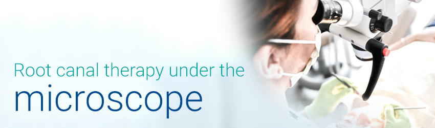
In such situations, canal therapy under the microscope is the only chance to cure such a tooth and save it from being extracted.
Owing to the magnification, many procedures can be performed with high efficiency. The performance of these procedures with a naked eye seems impossible or difficult, for instance,
- locating root canal orifices,
- determining the presence and locating additional canals,
- treatment of the root canal isthmus,
- filling root canals,
- monitoring the cleanliness of canal treatment,
- treatment of the perforation of the chamber floor and/or the root..
A microscope is also successfully used in other areas of dentistry:
- restorative dentistry (for instance, fillings, microtreatment of carious lesions),
- prosthetics (e.g., treatment of roots for posts, micro-preparations for veneers, restoration tightness check-ups),
- surgery (e.g., the possibility of using micro-instruments and thin sutures, thorough check-ups and diagnostics of tissues involved in the procedure),
- diagnostics (e.g., exposing leakage in existing fillings, initial caries lesions, lesions, enamel microcracks)